![]()

 |
|
 |
 At my last testing place, they foolishly gave me a CD with both my MRI and chest X-ray. It turns out the graphics are stored in some proprietary format. However, they put a viewer on the CD as well as the data. It took me just a couple of minutes to figure out how to run the viewer.
It turns out this viewer is browser based and runs inside my IE. When I was in the hospital and looked over my doctor’s shoulder at my CAT scans, I remember the software there was also browser based.
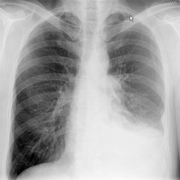
I don’t have the skill to read the MRI, but the chest X-ray is obvious to even to me.
You can see that my right lung looks great, but my left one looks half gone. My understanding is this is fluid between the lung and my rib cage. Looking at this photo, you can see why my aerobic capacity is restricted.
If you look closely, you may notice I accidentally also captured my cursor in the picture.
Posted by The Vorlon at October 17, 2005 7:37 PMI have no idea what I'm looking at either, but I can clearly see the area that contains the fluid.
I've had two MRIs recently. After the first I took the Xrays directly to my neurologist's office. When I got in the parking lot, I pulled them out and held them up to the light. There was one negative of my skull and brain. It had a huge white spot in it. I said to myself, "I don't think I want to look at these," and put them back in the envelope. As it turned out the doctor said everything was OK. As we aren't trained, it's difficult to look at anything and get a sense of the situation.
You mentioned in a previous entry that the lung had an inoperable tumor. Do you know if the fluid covering up the turmor? Also, by inoperable, I assume they mean inoperable at this time. Do they think the chemo will reduce the tumor to the point they can later operate? How are they attacking the fluid problem?
The xray is amazing. I go to the neurologist again on Friday. I think I'll grab my xray sheets and try to post a few scans myself. Having on CD is really nice.
Posted by: Reb Orrell at October 18, 2005 7:51 AM